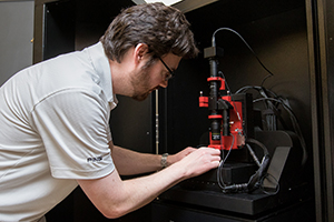
Laboratory Instrumentation
The Natural Sciences Laboratories and Field Facilities support practical learning
experiences that fully complement the science courses and academic programs offered
by the University.
Instrumentation
The Biology and Chemistry programs are housed mainly in the Unified Science Center where introductory and advanced laboratory courses in these disciplines are offered. Advanced instrumentation and research rooms are located within the Unified Science Center. Undergraduate teaching and research utilize the following instrumentation and equipment:
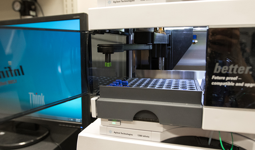
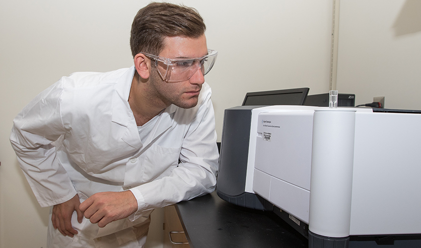
Agilent 1260 High Performance Liquid Chromatograph (HPLC) with diode array detector
A pressure range of up to 600 bar with a flow rate up to
5 mL/min allows the use of almost any column – conventional, sub-2 µm-particle or
superficially porous columns. A UV, fluorescence, or ELSD detector provides the data
rate required for high-resolution separations or fast analyses.
Molecular Devices Gene Pix 4100A
Genomics
- SNP genotyping arrays
- Arrayed comparative genomic hybridization (aCGH) arrays
Transcriptomics
- Gene expression arrays
- RNA interference (RNAi) arrays
- MicroRNA (miRNA) arrays
Proteomics
- Protein arrays
- Peptide arrays
- Antibody arrays
- ELISA arrays
- Reverse Phase Arrays
Epigenomics
- DNA methylation arrays
-
Chromatin immunoprecipitation (ChIP) arrays
Novel Applications
- Tissue arrays
- Quantum Dots
- Carbohydrate Arrays
- Chemical Compound Arrays
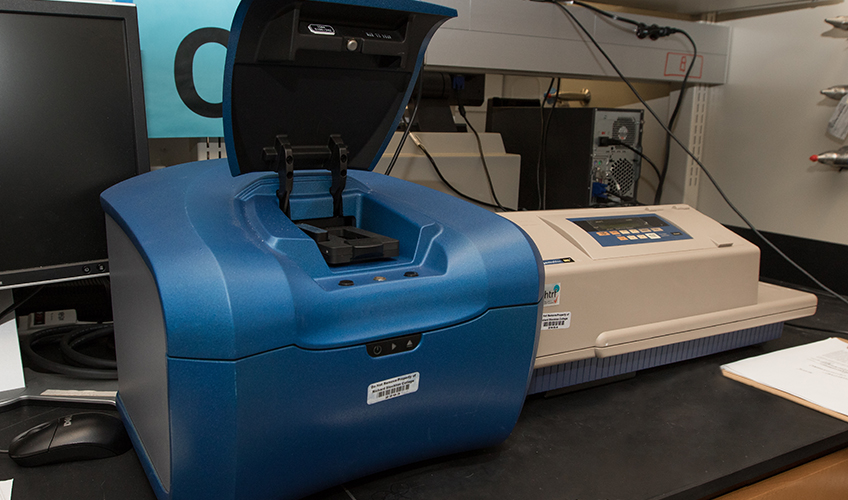
Spectramax M5 microplate reader Spectrophotometer
The optical systems use two scanning monochromators so the user can determine optimal
excitation and emission settings, resulting in assay performance similar to
that of dedicated single-mode readers.
Source: Molecular Devices: SpectraMax M Series Multi-Mode Microplate Readers Manual and Molecular Devices GenePix 4100A Microarray Scanner website
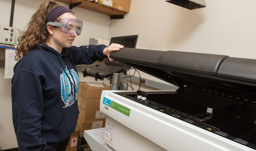
Perkin Elmer Tri Carb 3110TR Liquid Scintillation Analyzer
The Tri-Carb® 3110TR is a computer-controlled benchtop liquid scintillation analyzer for detecting small amounts of alpha, beta, and gamma radioactivity.
Source: PerkinElmer Tri-Carb 3110TR Low Activity Liquid Scintillation Analyzer Spec Sheet
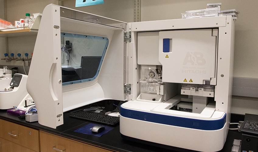
Applied Biosystems Hitachi 3500 Genetic Analyzer
The Applied Biosystems 3500/3500xL Genetic Analyzers are fluorescence-based DNA analysis instruments using capillary electrophoresis technology with 8- or 24-capillaries.
Source: Applied Biosystems 3500/3500XL Genetic Analyzer User Guide
Instrumentation used in the 2019 1st Place NAMS symposium: Analysis of Soil Bacteria for the Production of Antibiotics
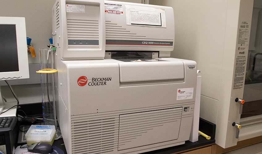
Beckman CEQ8000XL Automated DNA Sequencer
This system is fully automated and capable of determining the base sequence and fragment length of DNA samples that have been prepared with dye-labeled reagents. Four-color, dye-labeled terminator chemistry kits are used to process samples for base sequence analysis. Generation of samples for fragment length analysis is performed using dye-labeled primers.
Source: CEQTM 8000 Genetic Analysis System User's Guide
The Biomek® 3000 Laboratory Automation Workstation
The Biomek® 3000 Laboratory Automation Workstation is a multi-axis liquid handling instrument designed for benchtop use and to fit in a laminar flow or fume hood for sterile or hazardous operations. The open architecture design, along with the extensible operating software, provides a foundation for integrating current and future specific-use components.
The Biomek 3000 workstation is a single-head instrument with a series of interchangeable tools. Different tools provide options for performing a variety of functions, including liquid transfer and plate washing operations, and moving labware around the deck. The modular design of the Biomek 3000 workstation allows expansion of the instrument capabilities to include additional operations such as filtration, plate shaking, photometric microplate measurement, and high-capacity operation.


A variety of labware and hardware adapt the deck of the Biomek 3000 workstation to accomplish multiple tasks, ranging from performing simple labware positioning and liquid transfers to completing complex assays that typically require additional devices in the laboratory.
Source: Beckman Coutler Biomek® 3000 Laboratory Automation Workstation User's Manual
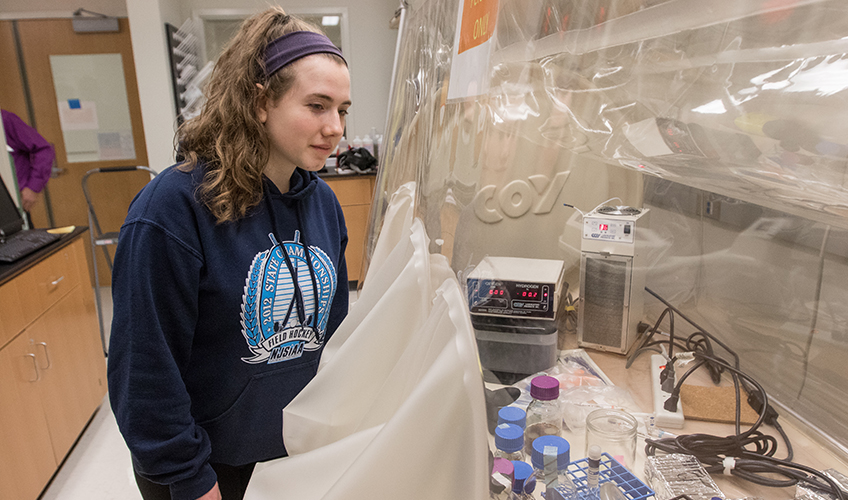
Coy Anaerobic Chamber
Used for anaerobic microbiology research, these units are also critical to many other research areas such as protein purification, clinical microbiology, biochemistry, cell culture, human microbiome studies, biofuels, and more.
Source: Coylab.com/products/anareobic-chambers
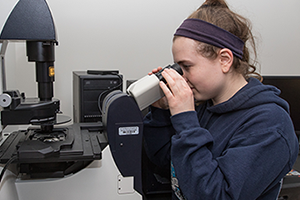
Leica Microsystems Microscope
Precise 5W LED illumination ideal for cell and tissue culture, microamplification, and live cell examinations.
Source: Leica Microsystems
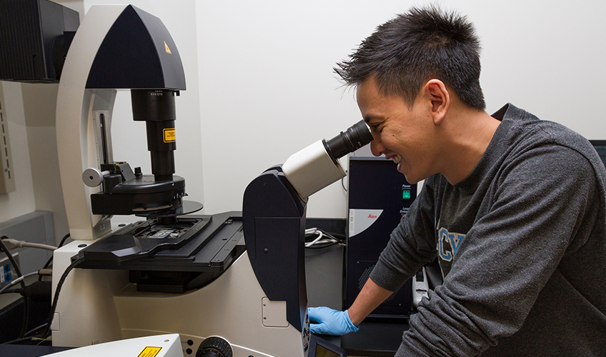
Leica TCS SPE Confocal Microscope
Providing all of the features needed for routine confocal techniques, it offers excellent quality imaging.
- The common Leica LAS X interface facilitates direct operation.
- Provides true spectral detection suitable for a lambda scan.
- It is intuitively controlled, and even users who are new to confocal microscopy will get publication-quality results immediately.
High Fidelity Spectral Imaging
The prism-based spectral detection – unique to all Leica confocal microscopes – and a highly dynamic photomultiplier offer extraordinary signal efficiency for gapless detection of even weak signals. A variety of robust solid-state lasers allows the use of a broad range of common dyes.
Source: Leica Microsystems
Molecular Devices Gene Pix 4100A
Genomics
Transcriptomics
Proteomics
Epigenomics
Novel Applications
Source: Moleculardevices.com
Spectramax M5 microplate reader Spectrophotometer
The optical systems use two scanning monochromators so the user can determine optimal
excitation and emission settings, resulting in assay performance similar to
that of dedicated single-mode readers.
Source: Spectramax M Series Multi-Mode Microplate Readers User Manual
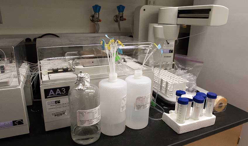
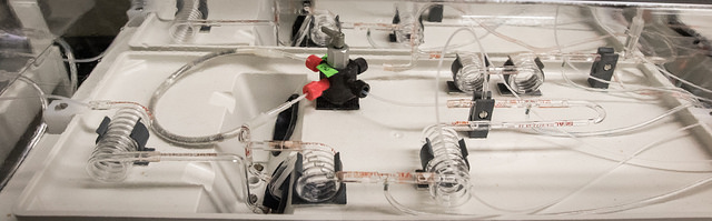
Seal Analytical AA3 Dissolved Nutrient Analyzer
Based on air Segmented Flow Analysis techniques (also known as Continuous Flow Analysis), the SEAL AutoAnalyzer 3 fully automates repetitive and complex sample analysis steps, from start-up to shutdown. The SEAL AA3 HR chemistry analyzer can also perform solvent extraction, distillation, gas diffusion, on-line filtration, and in-line UV digestion in a continuously flowing stream. SEAL AA3 HR chemistry analyzers are fully supported with a library of over 700 documented applications, including USEPA, ISO, and ASTM standard methods.
Source: SealAnalytical.com
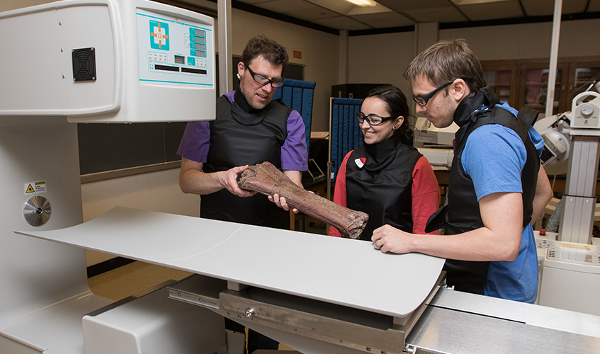
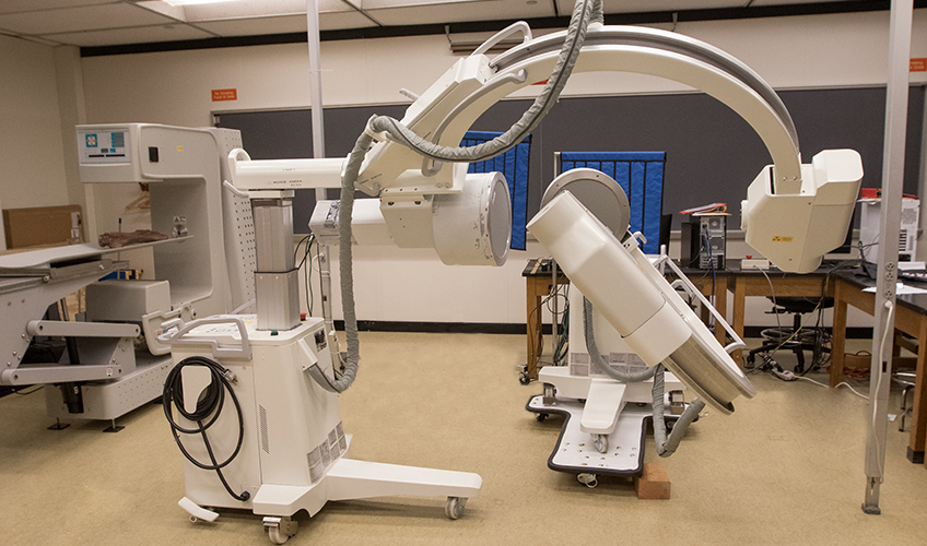
XROMM Analyzer
X-Ray Reconstruction of Moving Morphology (XROMM) is a 3D imaging technology for visualizing rapid skeletal movement in vivo.
XROMM combines 3D models of bone morphology with movement data from biplanar x-ray video
to create highly accurate (±0.1 mm) re-animations of the 3D bones moving in 3D space.
Rapid bone motion, such as during bird flight, frog jumping, and human running, can
be visualized and quantified with XROMM.
Source: Xromm.org
Agilent 1260 High Performance Liquid Chromatograph (HPLC) with diode array detector
A pressure range of up to 600 bar with a flow rate up to
5 mL/min allows the use of almost any column – conventional, sub-2 µm-particle or
superficially porous columns. A UV, fluorescence, or ELSD detector provides the data rate required for high-resolution
separations or fast analyses.
Source: labwrench.com
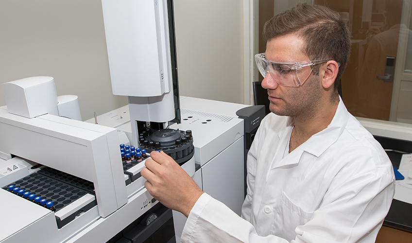
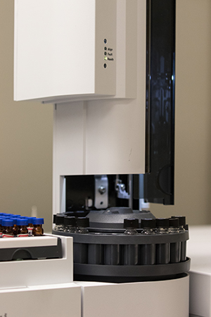
Agilent 7890B Gas Chromatographs (GC) with autosamplers and a variety of detectors: ECD, TCD, FID, MSD
Gas chromatography, coupled with static
headspace sampling is an easy-to-use,
high-throughput tool for determining
residual solvent impurities in pharmaceutical
products. Sample preparation is simple, and
the method is easily validated. In addition,
headspace sampling allows you to avoid
matrix injections that can cause column
degradation and coelution.
Source: Agilent 7890B Gas Chromatograph brochure
Instrumentation used in the 2019 1st Place NAMS symposium Analysis of Soil Bacteria for the Production of Antibiotics
Agilent Cary Eclipse Fluorescence Spectrophotometer
The fibre optic capabilities extend the utility of the system for simplified measurement of solid samples outside the sample compartment or for the direct measurement of cold, hot, or potentially dangerous samples, down to very low volumes.
Food
- Additives and supplements. Quality
Assurance testing and monitoring - Authenticity and food origin
- Food packaging and processing
including hydrocarbon contamination
through processing
Environmental
- Preliminary Identification of hydrocarbon
origin in oil spills using synchronous
scanning - Waste water tracing into environmental
flows by using fluorescent markers - Investigation of the source of organic
matter pollution in river and sea water
Chemical and Materials Applications
- Determination of fluorescent properties
of cleaning products - Investigation of the fluorescent
properties of optical components - Analysis of surface contamination by
fluorescent organic compounds in
manufacturing processes - Polarized (anisotropy) measurements
for understanding the molecular
environment during polymer research
Source: Agilent Cary Eclipse Fluorescence Spectrophotometer spec sheet
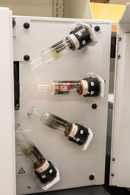
Agilent 240 FS AA Flame Atomic Absorption Spectrophotometer Hollow Cathode Lamps
Atomic absorption spectrophotometry analyzes the concentration of elements in a liquid sample based on energy absorbed from certain wavelengths of light (usually 190 to 900 nm). Atomic absorption spectrophotometers typically include a flame burner to atomize the sample (most commonly a hollow cathode lamp), a monochromator, and a photon detector. Equipped with a turret or fixed lamp socket that can hold multiple lamps (up to eight) to reduce downtime between samples or allow for sequential analysis.
Typical sensitivity for an atomic absorption spectrometer using a flame burner is in the parts per million range. For trace analysis, a graphite furnace can be used in place of a flame burner to increase the sensitivity by several orders of magnitude (in the parts per billion range). Atomic absorption spectrophotometers are used in many industries, including environmental testing, metal analysis, semiconductor manufacturing, petroleum and chemical production, and in pharmaceuticals, for example.
Source: labcompare.com
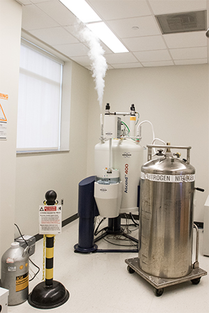
Bruker Advance 400 MHz Nuclear Magnetic Resonance (NMR) Spectrometer
Analytical nuclear magnetic resonance (NMR) solutions and instruments for life science and material research applications.
Nuclear magnetic resonance spectroscopy is used to study the structure of molecules, the interaction of various molecules, the kinetics or dynamics of molecules and the composition of mixtures of biological or synthetic solutions or composites.
The size of the molecules analyzed can range from a small organic molecule or metabolite, to a mid-sized peptide or a natural product, all the way up to proteins of several tens of kDa in molecular weight.
NMR nuclear spectroscopy complements other structural and analytical techniques such as X-ray, crystallography, and mass spectrometry. NMR’s advantage is the unique ability of a nuclear spectrometer to allow both the non-destructive and the quantitative study of molecules in solution and in solid state, as well as to enable the study of biological fluids.
Source: bruker.com
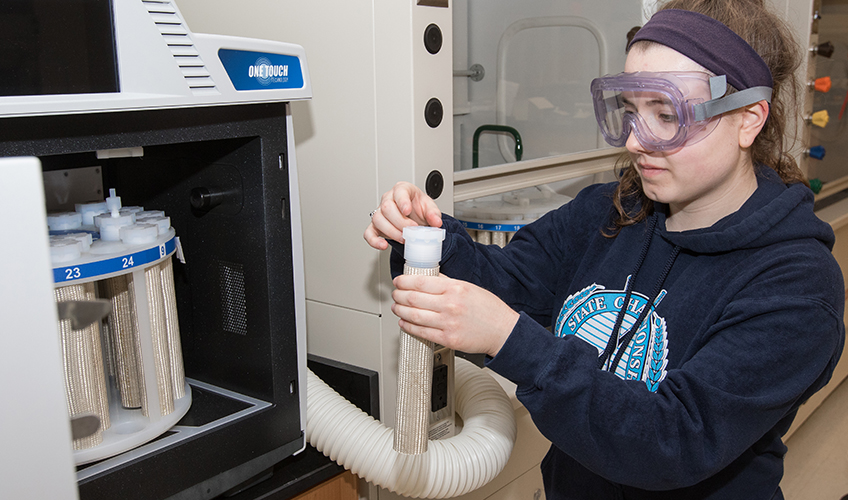
CEM MARS 6 Microwave system
Microwave is a technique used to dissolve solid sample matrices into an aqueous liquid. This is achieved by placing a sample in a concentrated acid matrix in a closed vessel and exposing it to microwave irradiation. Both the speed of thermal decomposition of the sample and the solubility of metals are increased. Once these metals are in solution, they can be quantified through spectroscopic techniques. The MARS 6 acid digestion process routinely cuts the time of sample preparation by 50 – 75 % as compared to hot plates and hot block.
Source: CEM.com
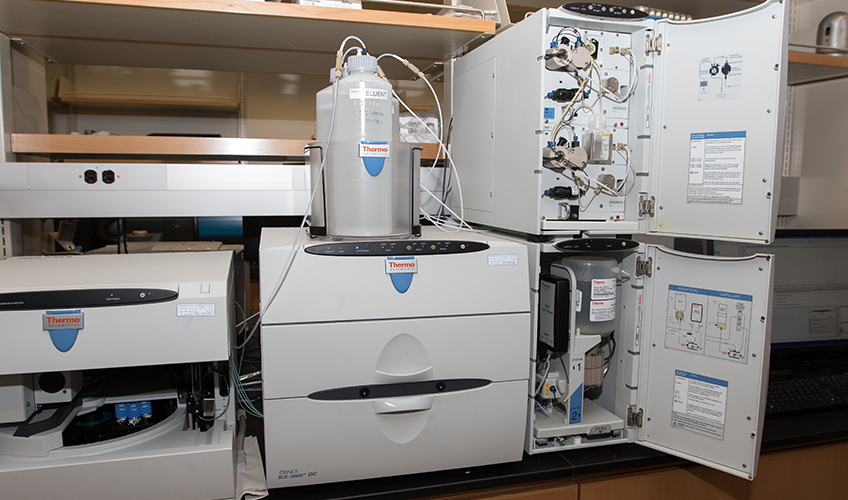
Dionex 5000 Ion Chromatographs (IC)
Ideally suited for trace level determinations with high matrix concentrations, using a standard or microbore column in the first dimension to separate analytes from matrix, followed by a capillary separation in the second dimension. This method provides high sensitivity, enabling conductivity detection limits to rival those of mass spectrometry.
Source: ThermoFisher.com
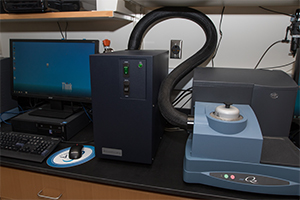
TA Instruments Q10 Differential Scanning Calorimeter (DSC)
The most common DSC application is the precise measurement of transition temperature.
Whether a melting temperature of a polymer
or the polymorphic transition of a pharmaceutical,
DSC provides the information quickly and easily on a minimum amount of sample. Important
temperature measurements include:
- Melting Temperature
- Glass Transition Temperature
- Thermal Stability Temperature
- Oxidation Onset Temperature
- Cure Onset Temperature
- Crystallization Temperature
- Polymorphic Transition Temperature
- Liquid Crystal Temperature
- Protein Denaturation Temperature
- Solid-Solid Transition Temperature
Source: tainstruments.com/brochure
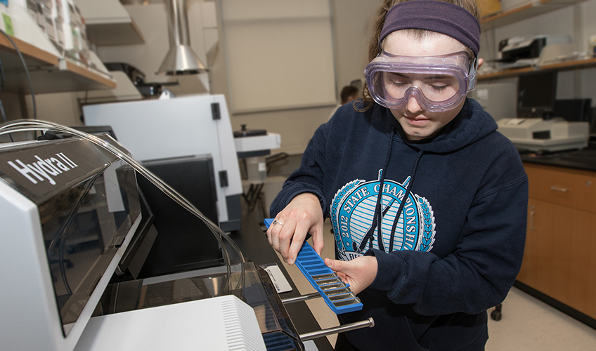
Teledyne Leeman Labs Hydra II Mercury Analyzer
It is a fully automated turnkey analyzer that measures mercury in solid and semi-solid sample matrices directly without any acid digestion (sample preparation). The system employs the technique of sample combustion (thermal decomposition), mercury concentration via gold amalgamation and detection by cold vapor atomic absorption.
Source: teledyneleemanlabs.com
Thermo Nicolet iS5 FTIR with ATR attachment
Infrared spectroscopy is the analytical technique for rapid identification of unknown materials (solids, liquids, and gases) and product quality screening.
Source: fishersci.com
Thermo Nicolet iS50 FTIR Spectrometer with ATR and multi-range beam splitter
The Nicolet iS50 analyzes complex mixtures and materials using its GC-IR or TGA-IR capabilities. The new OMNIC Mercury software automatically isolates and identifies compounds eluted from a GC column or evolved from a TGA. This functionality is especially useful in TGA, where several compounds can evolve at the same time. Such analyses provide insight into subtle formulation differences in plastics and rubbers, which can lead to not-so-subtle differences in product performance and quality.
Source: News-medical.net
Nicolet FT-IR Spectrometer
Uses infrared energy to excite the atoms of a material. The spectrum of the light absorbed and transmitted as the atoms vibrate provides a fingerprint of the structure of the molecules in the sample. This fingerprint can used to identify the chemical bonds present in the sample.
The Nicolet 6700 is currently equipped with a diamond crystal single bounce attenuated total reflectance (ATR) attachment
Rigaku MiniFlex 600 X-ray Diffractometer (powder) with 6-sample changer
MiniFlex X-ray diffractometer (XRD) is a multipurpose analytical instrument that can determine: phase identification and quantification, percent (%) crystallinity, crystallite size and strain, lattice parameter refinement, Rietveld refinement, and molecular structure. It is widely used in research, especially in material science and chemistry, as well as in industry for research and quality control.
Source: Rigaku.com
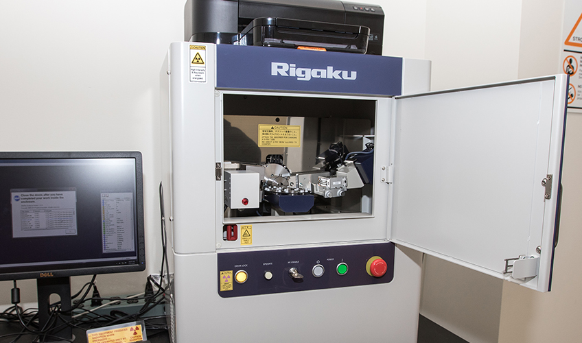
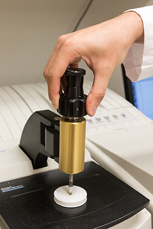
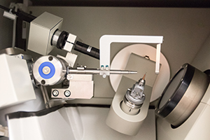
Rigaku XtaLAB mini X-ray Diffractometer (single crystal) (XRD)
The Rigaku XtaLAB, a benchtop X-ray crystallography system, is a compact single-crystal X-ray diffractometer designed to produce publication-quality 3D structures. Rapidly analyze new compounds as they are synthesized in the lab.
Source: Rigaku.com
Coming Soon
Coming Soon
NanoMagnetics Atomic Force Microscope (AFM)
Create images of atoms and map the surface structure materials. Offers Excellent features such as alignment-free design, closed-loop flexure scanner, decoupled z scanner, 10 MP video microscope, and flexible operating modes.
Source: Nanomagnetics Atomic Force Microscope Flyer
More Coming Soon
Coming Soon
Facilities
The School of Natural Sciences and Mathematics (NAMS) requires an extensive on-campus and off-campus infrastructure to accomplish its academic mission in the different scientific disciplines.
Most of the infrastructure is located on the Galloway campus in the Arts and Sciences (A&S) building, and the Academic Quad comprised of three newly constructed buildings (2013-2018): Unified Science Center 1 (USC1), Unified Science Center 2 (USC2) and the John F. Scarpa Health Sciences Center (HSC), which is home to the Sustainability Program. A short 15-minute drive from the Galloway campus, you will arrive at the NAMS Marine Field Station (MFS) that serves as a launch point for the University’s fleet of research vessels, as well as the site of the University’s Coastal Research Center (CRC). Other areas on campus that also support the mission of NAMS are the Arboretum, Observatory, Sustainability Farm, Greenhouses, Vivarium, and open spaces for forestry and marine field research.
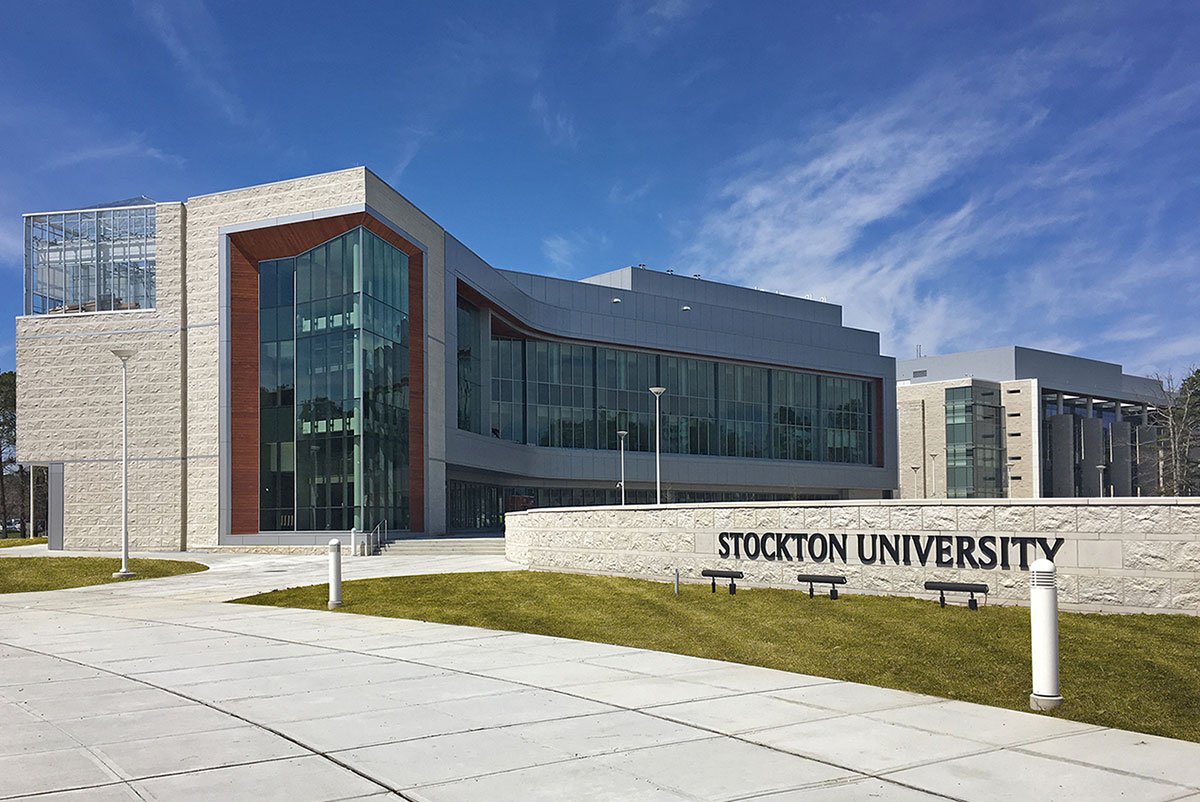
The Unified Science Center 2 (USC2) is a state-of-the-art facility that will allow Stockton to accommodate more students and better prepare them for successful careers in science, technology, engineering, and mathematics (STEM). The academic quad expansion will include the $33.2 million USC2 and a $15.2 million John F. Scarpa Health Sciences Center, near the existing Unified Science Center and Campus Center. The new buildings will be supported by funding from the Building Our Future Bond Act, which was overwhelmingly approved by New Jersey voters in 2012. The bond act will provide $21.465 million in funding for the USC2, and the University will pay 25 percent or $7.155 million. The John F. Scarpa Health Sciences Center will receive $13.5 million in funding from the bond issue, and the University will pay 25 percent or $4.5 million.
The main entrance of the academic quad will face Vera King Farris Drive and will provide a central location for students to study and meet between classes, as well as space for the University community to gather for campus events. The 58,210 thousand square foot Unified Science Center 2 will be an expansion to the existing 64,000-square-foot Unified Science Center. The three-story building will house teaching and research labs for various disciplines in the sciences, a vivarium, a large greenhouse, a multi-purpose room, and faculty offices. The 37,720-square-foot John F. Scarpa Health Sciences Center will include space for the Sustainability program, classrooms, faculty offices, and collaboration areas with tables and chairs.
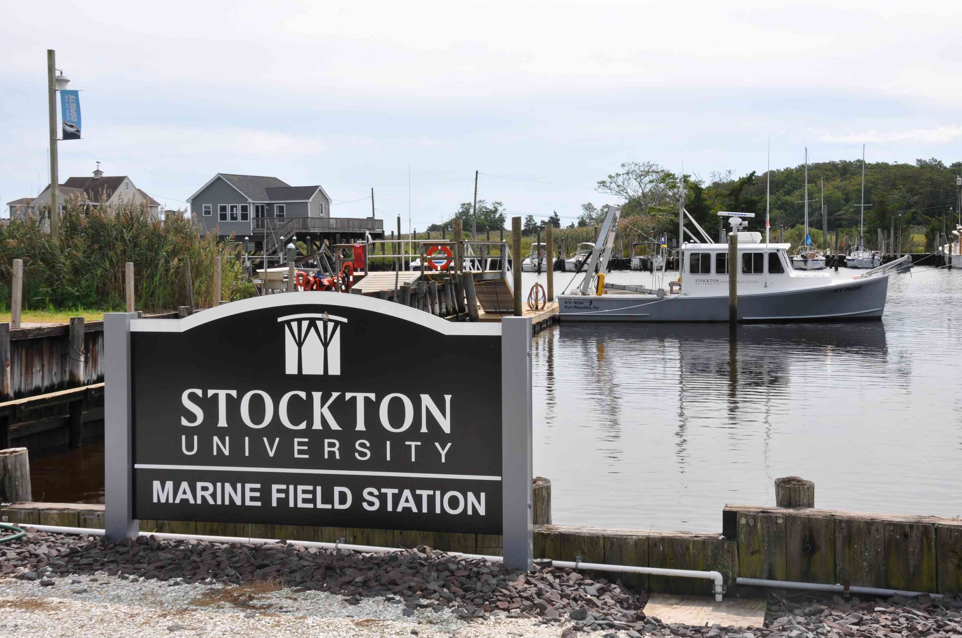
The Marine Field Station is located on an 8-acre parcel in Port Republic, New Jersey which is located within the Jacques Cousteau National Estuarine Research Reserve. It is conveniently located within 30 minutes of the Galloway and Atlantic City campuses – 15 minutes from Galloway and 30 minutes from the Atlantic City campus. The MFS is also the site of the University’s Coastal Research Center. The MFS offers laboratories and support facilities to provide access to an array of resources, including vessels, sampling equipment, and marine science technology instrumentation. The inshore research fleet consists of shallow-draft vessels ranging from 16’ – 24’ and the flagship vessel, the R/V Petrel, which is 36’ x 14’. The marine operations staff provide support services and oversee the maintenance and use of the facilities, vessels, instrumentation, and sampling equipment. The MFS is a University National Oceanographic Laboratory System (UNOLS) member and an organizational member of multiple professional societies, including the National Association of Marine Laboratories, the Organization of Biological Field Stations, Scientific Boating Safety Association, Marine Technology Society, and the Hydrographic Society of America.

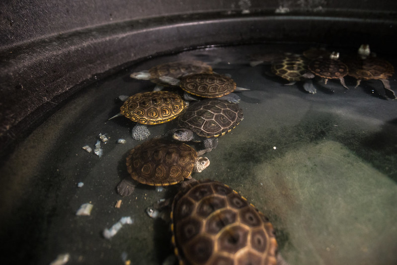
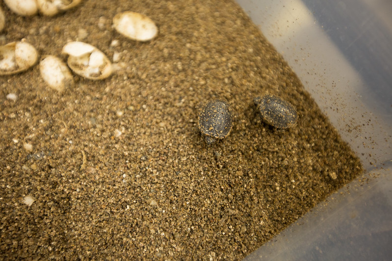
The Vivarium is located in USC2 and supports a variety of research and teaching display animals. One full-time and one part-time staff member oversee the 7-day-a-week care for the animals. The Vivarium is home to the terrapin project, which rehabilitates injured terrapins and also cares for mice under an NIH-funded research project. Many different species of rodents, fish, amphibians, and reptiles are also housed in this secured area, which is only accessible to properly trained faculty, staff, and students who have been trained in handling the various animals.
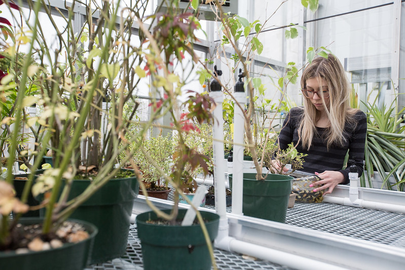
There are three Greenhouses located in different locations across the main campus (USC2, Arts & Sciences, and
the Sustainability Farm) and are used for both teaching and research. The greenhouses
are maintained by two staff members and student workers.
The USC2 greenhouse is the main location for the laboratory plant courses, which has
a large headhouse used for teaching students proper growing techniques. It houses
a large variety of plants used for teaching, research, and laboratory display. This
greenhouse is also used in collaboration with other schools within the university,
as well as for outreach events. The A&S greenhouse is a small addition to the building
used for soil analysis within the geology and environmental science classes. The Sustainability
Farm on campus has a large mobile greenhouse that allows a variety of plants to be
grown in a unique environment. This is a great experimental area for teaching and
research, which also extends the opportunity for off-season growing. The Sustainability
greenhouses are used extensively for organic research and laboratory activities.
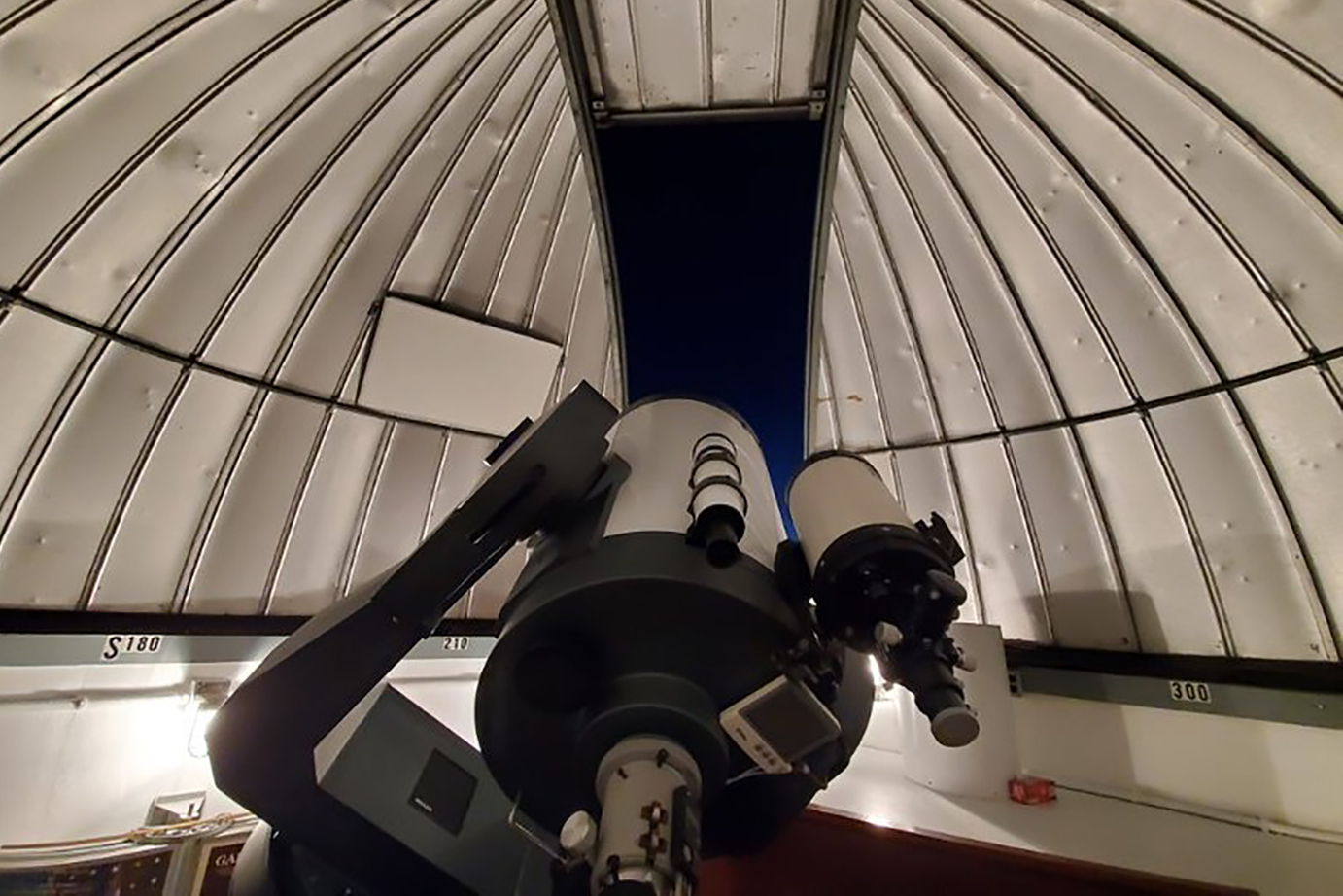
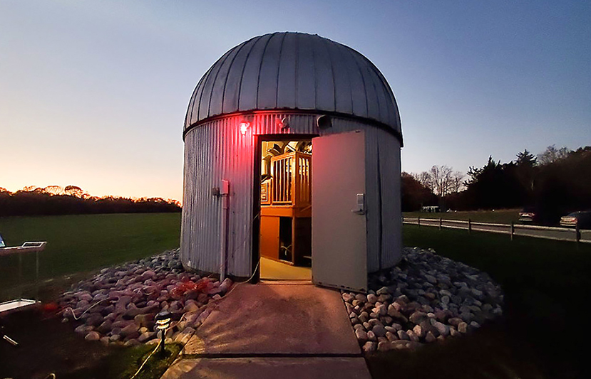
The Harold E. Taylor Observatory, located off Pomona Road, on the west campus, houses a 0.4 m Meade computer-controlled reflector telescope. It also contains two computer-controlled, portable reflector telescopes (8-inch and 6-inch). The Observatory is used for teaching General Astronomy courses to an average of 70 students per semester, and a hybrid Astrophysics course. It is also used for research and outreach programs. Observation nights are open to the Stockton community and the general public and are held approximately twice a month, weather permitting. For more information, please visit the observatory website.
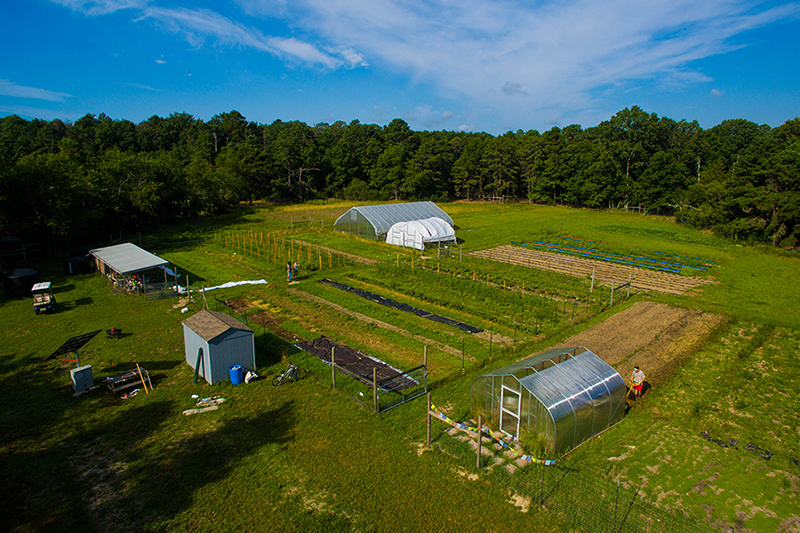
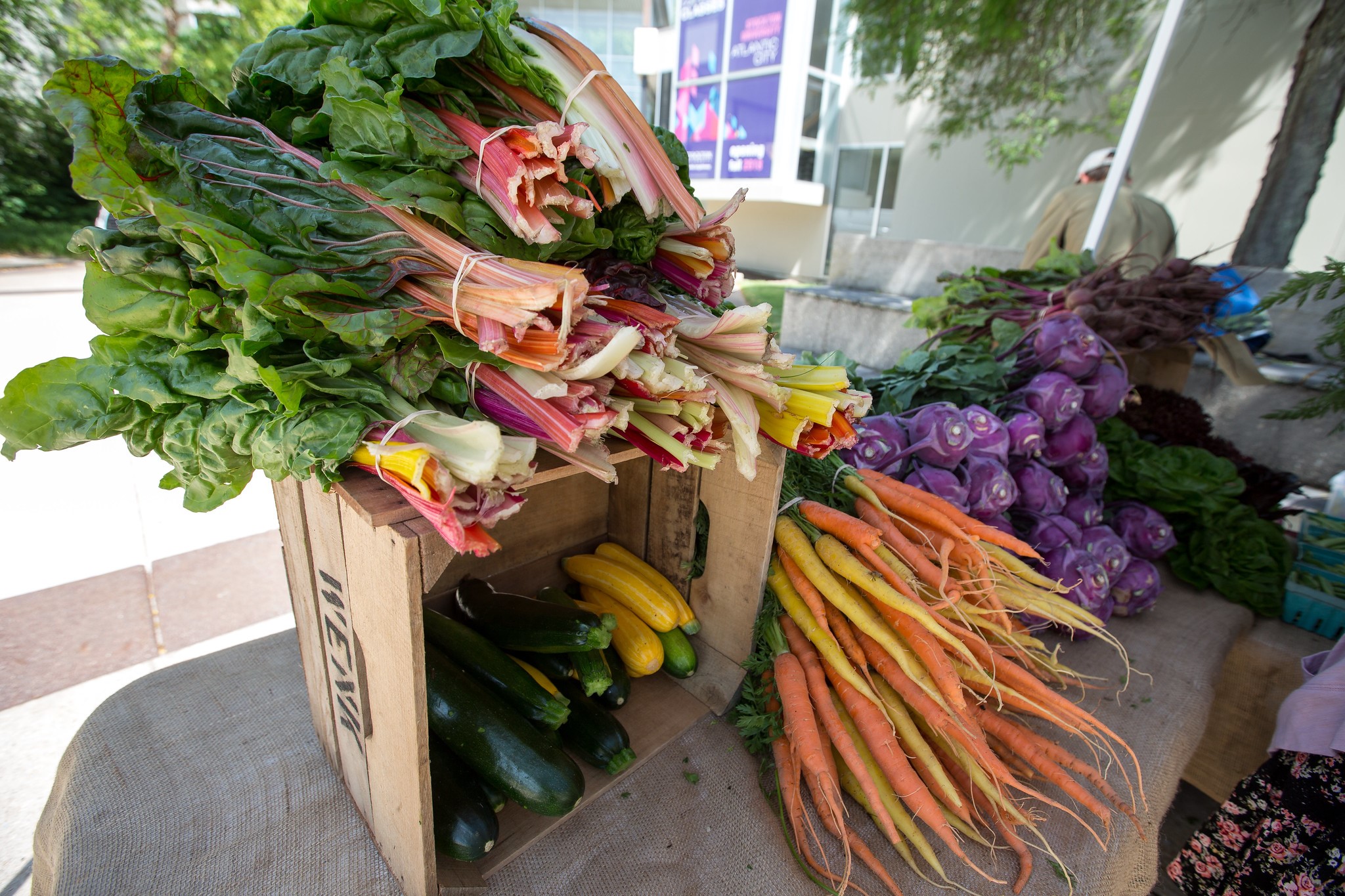
The Sustainability Farm was established in 2012 as a student-run project. The farm productivity and campus visibility have increased significantly relative to previous years. The infrastructure expanded to include a 30’x48’ high tunnel-greenhouse, increased water storage, solar power and storage, and a new fence, which increased the production area from one-third of an acre to 1.5 acres. A new structure for student involvement was introduced; students can now enroll in a Farm Practicum each semester, which ensures their commitment and formalizes the academic aspect of farm participation. Enrolled students produce and sell organic crops at the weekly farmers market on and off campus. The farm also has a large mobile greenhouse structure that can be moved to create different environments throughout the seasons and allows for the extension of the growing season for a variety of crops. Students practice hands-on training in sustainable agriculture through the use of sustainable practices such as solar and geothermal energy use, rainwater collection-based irrigation, and sustainable farming methods. For more information, please visit the farm website.